Cranial Cruciate Ligament Injury
What is the cranial cruciate ligament?
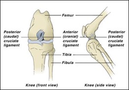
The cranial cruciate ligament (CrCL) in dogs is the equivalent to the anterior cruciate ligament (ACL) in humans. The cranial cruciate ligament is one of many ligaments in the dog’s knee, connecting the femur (thigh bone) to the tibia (shin bone) in their back legs. It has many functions, including preventing the shin bone (tibia) from sliding too far forward relative to the thigh bone (femur)

What are signs of a CrCL injury or rupture?
The signs will vary depending on how severe the ligament rupture is and when the injury occured. Partial rupture of the ligament is more subtle and can progress into complete ligament rupture.
- Non-weight bearing or tip-toeing on their hindlimb
- Abnormal posture
- Swelling and warmth in the knee
- Hindlimb sticking out to one side when sitting down
- Reluctance to rise, jump or walk
Signs of chronic rupture of the CrCL ligament:
- Lameness that has been persisting despite resting
- Slow, stiff gait
- Reluctance to rise, jump or walk
- Thickened tissue around the knee
- Abnormal posture
How is a CrCL injury diagnosed?
The diagnosis of a CrCL rupture is usually based on clinical signs, physical examination (e.g. a positive “cranial drawer sign”), and knee x-rays.
The cranial drawer motion can be performed when your dog is lying down on their side. Your vet will examine the shin bone and manipulate it gently to check if it moves forward when compared to the thigh bone. It is necessary to repeat this test under heavy sedation or general anaesthetic because most dogs are usually too painful and tense in the consult room for it to be performed correctly.
Additionally, x-rays of the knee will need to be performed under heavy sedation or general anaesthetic. While x-rays may not necessarily show the thick fibres of the CrCL (which would require an MRI), x-rays can be very helpful showing how the body reacts to a ligament injury such as increased swelling and inflammation within the joint, and evidence of arthritis. It can also help indicate other potential causes of hindlimb lameness.
It is absolutely necessary for the x-rays to be taken under heavy sedation or general anaesthetic due to a few reasons:
- A dog in pain will not be able to bend their leg accordingly
- Precise accurate x-rays are needed – this is required to make measurements and plan for any potential knee surgery. These x-rays cannot be taken if the dog is moving around or blurry.
Why does the CrCL rupture?
The rupture of the CrCL ligament is due to weakening of the ligament over time. Factors that may play a contributing role include abnormal hindlimb conformation, genetics, and obesity. The weak ligament is then ruptured following running or jumping.
Certain breeds are predisposed to a CrCL ligament rupture, but any breed can be affected. However, we see CrCL injury most commonly in:
- Labrador Retriever
- Golden Retriever
- Rottweiler
- German Shepherd
- Bernese Mountain Dog
- Mastiff
- Saint Bernard
- French Bulldog
What is the treatment for CrCL rupture?
A specialist orthopaedic surgery is the recommended treatment for cranial cruciate ligament (CrCL) ruptures in dogs.
There are three different types of surgical techniques available.
- Tibial plateau levelling osteotomy (TPLO). This is a commonly recommended technique by orthopaedic specialist surgeons due to the high success rate and eligibility of most patients. This technique involves making a circular cut around the top of the shin bone and rotating it until it achieves a near level orientation that minimises sliding forces, resulting in a biomechanically stable joint without the need for a cranial cruciate ligament (CrCL). Simply put, think of a wagon on a slope tied to a post with a piece of rope (equivalent to the CrCL) holding it in place so the wagon doesn’t slide down hill. The TPLO procedure changes the wagon to level ground so the piece of rope is no longer required for the wagon to remain stable or still, even with extra weight loaded on.
- Tibial tuberosity advancement (TTA). Similar to a TPLO, a biomechanically stable joint is created with a linear cut along the front of the shin bone and advancing it forward until a near level orientation is achieved.
- Extracapsular repair. This involves using suture material placed under the skin and just outside the knee joint to mimic the function of the CrCL.
If you would like to see a model of the TPLO surgical technique, watch this video:
TPLO thrust logo from Fitzpatrick Referrals on Vimeo.
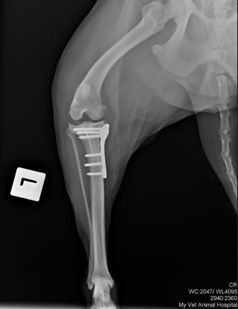
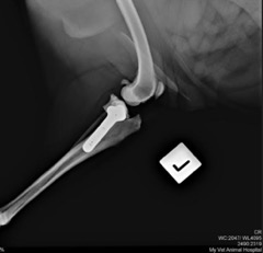
These are x-rays of a patient’s knee who had TPLO surgery at My Vet Animal Hospital:


What is involved in the recovery period for CrCL surgery?
Your vet will guide you through a comprehensive schedule – depending on which surgical technique has been used.
Here at My Vet Animal Hospital, in addition to staying the entire day of surgery, our patients return the following day for further close monitoring. From there, weekly check ups are performed to ensure your furbaby is recovering as expected until 6 weeks post-op when repeat x-rays are performed.
As a quick overview, your furbaby’s recovery period would involve:
- Day 0: Surgery + full day hospitalisation and monitoring
- Day 1 post-op: Full day hospitalisation
- Day 3 post-op: Recheck with a vet
- Week 1 post-op: Recheck with a vet
- Week 2 post-op: Recheck with a vet + suture removal + 1st joint support/arthritis injection (Zydax) + commence physiotherapy
- Week 3 post-op: 2nd joint support/arthritis injection (Zydax)
- Week 4 post-op: 3rd joint support arthritis injection (Zydax)
- Week 5 post-op: 4th joint support/arthritis injection (Zydax)
- Week 6 post-op: Repeat x-rays
At home, it is important that your furbaby has the following:
- Exercise restriction. This means CONFINEMENT to a small area (e.g. 2 metres by 2 metres) so it is definitely worth investing in a doggy fence or a playpen. Once exercise can be slowly re-introduced, low-impact activities are recommended like swimming.
- Avoid stairs, slippery floors and any form of jumping
- Daily medication. These are often given orally every day and include pain relief/anti-inflammatory medications for the first 4-6 weeks as well as antibiotics for the first 2 weeks. Monitor closely for any side effects such as gut upsets
- Keep the surgical site dry and clean
- Commence physiotherapy exercises from 2 weeks post-op
- Commence daily arthritis supplementation long-term
- Dietary changes – especially if your pet is overweight! We recommend considering switching to a prescription diet specifically for joint support as well as weight loss like Hill’s metabolic + mobility
What are the complications associated with surgery?
Complication rates are very rare with experienced specialist orthopaedic surgeons, so that is why we always recommend having a board-certified specialist veterinary surgeon perform the procedure. We have a great existing relationship with Consulting Animal Specialists which allows us to organise certain complex surgical procedures to be performed by visiting specialists out of the comfort and convenience of our clinic.
Some of the common surgical complications may include:
- Infection
- Implant rejection
- Implant problems if your dog is not rested properly in the first crucial 6 weeks after surgery
It is important to note that up to 50% of dogs who rupture their CrCL ligament will tear the other side within 2 years of each other.
What happens if I don’t elect to have surgery performed?
Once the cranial cruciate ligament ruptures, it cannot repair itself.
If left untreated, it will cause:
- Chronic instability in the knee joint
- Chronic pain and inflammation in the hindlimb
- Further damage to supporting structures in the knee joint such as the cartilaginous cushion (meniscus) that acts as a shock absorber
- Irreversible osteoarthritis changes
- Loss of muscle mass in hindlimb
- Reluctance to go on walks or runs
- Poor quality of life
What is the prognosis after CrCL surgery?
Prognosis is very good with surgery to repair cruciate ligament ruptures in dogs as long as the proper steps are taken during the recovery period. Around 90-95% of dogs with TPLO or TTA surgery will return to 95-100% normal function.
At My Vet Animal Hospital, we are focused on providing the highest quality care to our patients. We work closely with orthopaedic surgeons who will perform the surgical procedure in the comfort of our vet hospital (your dog will be happier with people that he/she knows too!). Many pet owners also prefer this as it is closer to their homes which makes follow-up appointments easier and less stressful for their dogs.


